How Can You Treat Bursitis Of The Foot
Overview
Retrocalcaneal bursitis and Achilles bursitis are the most widely spread types of ankle / heel bursitis out there. However, there are several bursa lubrication fluid sacs behind the heel bone protecting this area that may become irritated, inflammed and painful.
Causes
Pain at the posterior heel or ankle is most commonly caused by pathology at either the posterior calcaneus (at the calcaneal insertion site of the Achilles tendon) or at its associated bursae. Two bursae are located just superior to the insertion of the Achilles (calcaneal) tendon. Anterior or deep to the tendon is the retrocalcaneal (subtendinous) bursa, which is located between the Achilles tendon and the calcaneus. Posterior or superficial to the Achilles tendon is the subcutaneous calcaneal bursa, also called the Achilles bursa. This bursa is located between the skin and posterior aspect of the distal Achilles tendon. Inflammation of either or both of these bursa can cause pain at the posterior heel and ankle region.
Symptoms
The main symptom of heel bursitis is pain. You may experience pain in your heel when you walk or run. There may also be pain if the area is touched or if you stand on your tiptoes. In addition to pain, the area may appear red and warm, which are both signs of inflammation. Even if you have these symptoms, only a doctor can determine if you have bursitis of the heel. Your doctor will use these symptoms along with a general exam to determine if you are suffering from bursitis of the heel.
Diagnosis
The doctor will discuss your symptoms and visually assess the bones and soft tissue in your foot. If a soft tissue injury is suspected, an MRI will likely be done to view where and how much the damage is in your ankle. An x-ray may be recommended to rule out a bone spur or other foreign body as the cause of your ankle pain. As the subcutaneous bursa is close to the surface of the skin, it is more susceptible to septic, or infectious, bursitis caused by a cut or scrape at the back of the heel. Septic bursitis required antibiotics to get rid of the infection. Your doctor will be able to determine whether there is an infection or not by drawing a small sample of the bursa fluid with a needle.
Non Surgical Treatment
Many cases of retrocalcaneal and retroachilles bursitis can be treated effectively at home. One of the most important factors is eliminating shoe gear that presses against the back of the heel. Comfortable, supportive footwear and frequently resting the foot will minimize friction at the heel and give the inflammation a chance to subside. These steps, along with other methods to alleviate swelling, such as non-steroidal anti-inflammatory drugs (NSAIDs, e.g., ibuprofen), icing the heel, and elevating the foot, are usually successful in treating retrocalcaneal bursitis.
Surgical Treatment
Bursectomy is a surgical procedure used to remove an inflamed or infected bursa, which is a fluid-filled sac that reduces friction between tissues of the body. Because retrocalcaneal bursitis can cause chronic inflammation, pain and discomfort, bursectomy may be used as a treatment for the condition when it is persistent and cannot be relived with other treatments. During this procedure, a surgeon makes small incisions so that a camera may be inserted into the joint. This camera is called an arthroscope. Another small incision is made so that surgical instruments can be inserted to remove the inflamed bursa.
Hammer Toe Pain After Surgery
 Overview
When a person has Hammer toes, the end of their toe bends downward and the middle joint curls up. Eventually, the toe gets stuck in a stiff, claw-like position. When the inside of your shoe rubs against a hammer toe, corns, blisters or calluses may form on top of the toe or on the bottom of your foot. This can make walking painful. You may also have pain in the joint where your big toe joins your foot. Hammer toe usually affects a person?s second toe (the toe next to the big toe), but it can affect other toes too.
Causes
Shoes that narrow toward the toe may make your forefoot look smaller. But they also push the smaller toes into a flexed (bent) position. The toes rub against the shoe, leading to the formation of corns and calluses, which further aggravate the condition. A higher heel forces the foot down and squishes the Hammer toes toes against the shoe, increasing the pressure and the bend in the toe. Eventually, the toe muscles become unable to straighten the toe, even when there is no confining shoe.
 Symptoms
A hammertoe may be present but not always painful unless irritated by shoes. One may have enlarged toe joints with some thickened skin and no redness or swelling. However, if shoes create pressure on the joint, the pain will usually range from pinching and squeezing to sharp and burning. In long standing conditions, the dislocated joints can cause the pain of arthritis.
Diagnosis
First push up on the bottom of the metatarsal head associated with the affected toe and see if the toe straightens out. If it does, then an orthotic could correct the problem, usually with a metatarsal pad. If the toe does not straighten out when the metatarsal head is pushed up, then that indicates that contracture in the capsule and ligaments (capsule contracts because the joint was in the wrong position for too long) of the MTP joint has set in and surgery is required. Orthotics are generally required post-surgically.
Non Surgical Treatment
A person with hammer toes will be asked to practice some exercises for their toes to regain average structure and movement. The exercises usually involve stretching and strengthening their toes. The person may attempt to pick things up off the floor using only their toes. They may also stretch their toes on a regular basis by hand to ease them into straightening out. Another example of a physical exercise specifically for a person's toes involves crumpling a towel with the toes. The towel can lie underneath the person's feet and the person can use their toes to scrunch up the towel as they perform simple tasks such as reading a book or watching television.
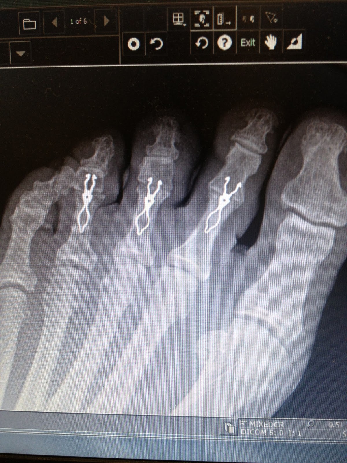
Surgical Treatment
Hammertoe surgery is performed when conservative measures have been exhausted and pain or deformity still persists. The surgery is performed on an outpatient basis. It typically required about one hour of time. An incision is placed over the inter-phalangeal joint. Once the bone is exposed, the end portion of the bone is removed. Your surgeon may then use pins or other fixation devices to assist in straightening the toe. These devices may be removed at a later date if necessary. Recovery for hammertoe surgery is approximately 10 to 14 days. You are able to walk immediately following the surgery in a surgical shoe. Swelling may be present but is managed as needed. Physical therapy is used to help reduce swelling in the toe or toes after surgery. Most of these toe surgeries can be performed in the office or the outpatient surgery under local anesthesia.
Is Over-Pronation Of The Feet Uncomfortable
Overview
Overpronation is a condition in which the foot rolls excessively down and inward. The arch may elongate and collapse (or ?fall?) and the heel will lean inward. Overpronation should not be confused with pronation. Pronation is a normal motion of the foot during weight bearing and allows the foot to absorb shock as it contacts the ground.
Causes
Unless there is a severe, acute injury, overpronation develops as a gradual biomechanical distortion. Several factors contribute to developing overpronation, including tibialis posterior weakness, ligament weakness, excess weight, pes planus (flat foot), genu valgum (knock knees), subtalar eversion, or other biomechanical distortions in the foot or ankle. Tibialis posterior weakness is one of the primary factors leading to overpronation. Pronation primarily is controlled by the architecture of the foot and eccentric activation of the tibialis posterior. If the tibialis posterior is weak, the muscle cannot adequately slow the natural pronation cycle.
Symptoms
It is important to note that pronation is not wrong or bad for you. In fact, our feet need to pronate and supinate to achieve proper gait. Pronation (rolling inwards) absorbs shock and supination (rolling outwards) propels our feet forward. It is our body?s natural shock-absorbing mechanism. The problem is over-pronation i.e. the pronation movement goes too deep and lasts for too long, which hinders the foot from recovering and supinating. With every step, excess pronation impedes your natural walking pattern, causing an imbalance in the body and consequent excessive wear and tear in joints, muscles and ligaments. Some common complaints associated with over-pronation include Heel Pain (Plantar Fasciitis) ,Ball of foot pain, Achilles Tendonitis, Shin splints, Knee Pain, Lower Back Pain.
Diagnosis
A quick way to see if you over-pronate is to look for these signs. While standing straight with bare feet on the floor, look so see if the inside of your arch or sole touches the floor. Take a look at your hiking or running shoes; look for wear on the inside of the sole. Wet your feet and walk on a surface that will show the foot mark. If you have a neutral foot you should see your heel connected to the ball of your foot by a mark roughly half of width of your sole. If you over-pronate you will see greater than half and up to the full width of your sole.
Non Surgical Treatment
Wear shoes with straight or semicurved lasts. Motion-control or stability shoes with firm, multidensity midsoles and external control features that limit pronation are best. Over-the-counter orthotics or arch supports can help, too. You know you are making improvements when the wear pattern on your shoes becomes more normal. Overpronation causes extra stress and tightness to the muscles, so do a little extra stretching.
Surgical Treatment
The MBA implant is small titanium device that is inserted surgically into a small opening between the bones in the hind-mid foot: the talus (ankle bone) and the calcaneus (heel bone). The implant was developed to help restore the arch by acting as a mechanical block that prevents the foot from rolling-in (pronation). In the medical literature, the success rate for relief of pain is about 65-70%. Unfortunately, about 40% of people require surgical removal of the implant due to pain.
Physical Therapy And Severs Disease
Overview
Sever's Disease, also known as calcaneal apophysitis, is a disease of the growth plate of the bone and is characterized by pain in the heel of a child's foot, typically brought on by some form of injury or trauma. This condition is most common in children ages 10 to 15 and is frequently seen in active soccer, football, or baseball players. Sport shoes with cleats are also known to aggravate the condition. The disease mimics Achilles tendonitis, an inflammation of the tendon attached to the back of the heel. A tight Achilles tendon contributes to Sever's Disease by pulling excessively on the growth plate of the heel bone (calcaneus). Treatment includes cutting back on sports activities, calf muscle stretching exercises, heel cushions in the shoes, icing, and/or anti-inflammatory medications. Note: Please consult your physician before taking any medications.
Causes
Sever's disease usually develops as a result of overuse and is common in active children between the ages of 8 to 12. Activities that involve running or jumping can cause undue stress on the calcaneal apophysis. This in turn leads to the development of microscopic damage to the calcaneal apophysis resulting in inflammation and pain. Poor flexibility of the calf muscles and of the Achilles tendon, overpronation (feet rolled in) and inappropriate footwear are some of the other factors that can cause Sever's disease.
Symptoms
The main symptom of sever's disease is pain and tenderness at the back of the heel which is made worse with physical activity. Tenderness will be felt especially if you press in or give the back of the heel a squeeze from the sides. There may be a lump over the painful area. Another sign is tight calf muscles resulting with reduced range of motion at the ankle. Pain may go away after a period of rest from sporting activities only to return when the young person goes back to training.
Diagnosis
Sever?s disease is diagnosed based on a doctor?s physical examination of the lower leg, ankle, and foot. If the diagnosis is in question, the doctor may order x-rays or an MRI to determine if there are other injuries that may be causing the heel pain.
Non Surgical Treatment
It is important that those with Sever?s Disease are treated by a medical professional to reduce pain and allow children to continue to participate in sporting activities. The Athlete?s Foot recommends a visit to your local medical professional to be diagnosed correctly and receive specialised care. Symptoms include pain through the back of the heel where the Achilles tendon inserts into the heel bone, pain during activity especially running and jumping and the back of the heel may be tender to touch.
Prevention
To prevent Sever's Disease, fit your child with kid's shoes with good cushioning in the footbed, shock absorption in the heel, and support in the outsole. Make sure children wear supportive shoes, especially when they're running and jumping, to reduce the impact on the heel and strain on the developing bone and muscle structure of a kids' feet. Children's arch supports and heel cups comfortably support the foot and encourage healthy alignment while your child runs or walks.
What Causes Flat Feet In Adults?
Overview
A painful flat foot, or adult acquired flatfoot deformity, is a progressive collapsing of the arch of the foot that occurs as the posterior tibial tendon becomes insufficient due to various factors. Early stages may present with only pain along the posterior tibial tendon whereas advanced deformity usually results in arthritis and rigidity of the rearfoot and ankle. 
Causes
Flat footedness, most people who develop the condition already have flat feet. With overuse or continuous loading, a change occurs where the arch begins to flatten more than before, with pain and swelling developing on the inside of the ankle. Inadequate support from footwear may occasionally be a contributing factor. Trauma or injury, occasionally this condition may be due to fracture, sprain or direct blow to the tendon. Age, the risk of developing Posterior Tibial Tendon Dysfunction increases with age and research has suggested that middle aged women are more commonly affected. Other possible contributing factors - being overweight and inflammatory arthritis.
Symptoms
In many cases, adult flatfoot causes no pain or problems. In others, pain may be severe. Many people experience aching pain in the heel and arch and swelling along the inner side of the foot.
Diagnosis
Looking at the patient when they stand will usually demonstrate a flatfoot deformity (marked flattening of the medial longitudinal arch). The front part of the foot (forefoot) is often splayed out to the side. This leads to the presence of a ?too many toes? sign. This sign is present when the toes can be seen from directly behind the patient. The gait is often somewhat flatfooted as the patient has the dysfunctional posterior tibial tendon can no longer stabilize the arch of the foot. The physician?s touch will often demonstrate tenderness and sometimes swelling over the inside of the ankle just below the bony prominence (the medial malleolus). There may also be pain in the outside aspect of the ankle. This pain originates from impingement or compression of two tendons between the outside ankle bone (fibula) and the heel bone (calcaneus) when the patient is standing.
Non surgical Treatment
Nonoperative treatment of stage 1 and 2 acquired adult flatfoot deformity can be successful. General components of the treatment include the use of comfort shoes. Activity modification to avoid exacerbating activities. Weight loss if indicated. Specific components of treatment that over time can lead to marked improvement in symptoms include a high repetition, low resistance strengthening program. Appropriate bracing or a medial longitudinal arch support. If the posterior tibial tendon is intact, a series of exercises aimed at strengthening the elongated and dysfunctional tendon complex can be successful. In stage 2 deformities, this is combined with an ankle brace for a period of 2-3 months until the symptoms resolve. At this point, the patient is transitioned to an orthotic insert which may help to support the arch. In patients with stage 1 deformity it may be possible to use an arch support immediately. 
Surgical Treatment
If initial conservative therapy of posterior tibial tendon insufficiency fails, surgical treatment is considered. Operative treatment of stage 1 disease involves release of the tendon sheath, tenosynovectomy, debridement of the tendon with excision of flap tears, and repair of longitudinal tears. A short-leg walking cast is worn for 3 weeks postoperatively. Teasdall and Johnson reported complete relief of pain in 74% of 14 patients undergoing this treatment regimen for stage 1 disease. Surgical debridement of tenosynovitis in early stages is believed to possibly prevent progression of disease to later stages of dysfunction.
Reflexology Pain In Arch Of Right Foot
Overview
Fallen arches, also known as flat feet or pes planus, may be present at birth or develop later in life. The midfoot normally exhibits a slight arch, keeping this region raised during walking. Absence of the normal arch causes flattening of the sole. Many people with fallen arches have no associated symptoms, while others experience foot pain or fatigue. Treatment for fallen arches depends on the severity of the condition and associated symptoms.

Causes
There are a number of possible causes for arch pain, but the most common are structural imbalances of the foot, such a pronated foot (rolls inward at the ankles). This is often not enough in isolation to cause the problem, but in combination with other factors arch pain may develop. These other factors are usually associated with overuse - running, walking, hard surfaces and/or OFAD (on feet all day), usually combined with inadequate or non-supportive footwear. The more common specific causes of arch pain can be plantar fasciitis (strain of the plantar fascia - a strong ligament that supports the arch), tarsal tunnel syndrome (a pinched nerve at the ankle that refers pain to the arch), foot strain from a pronated foot or flat foot, there can be osteoarthritis of the joints in the midfoot that can cause arch pain.
Symptoms
Bones and ligaments work together to form joints, and bones are joined together by ligaments. Strains occur in ligaments. In the arch, there are ligaments that are located at the ends of each bone. These ligaments connect the bones to other bones on both ends and on the sides. Point tenderness and looseness of a joint are indicators of a sprain. Fractures are indicated by point tenderness that may be severe over the area of bone that is affected. There may be a distinguishable lump or gap at the site of the fracture. A rotated toe or forefoot may also be a sign of a fracture.
Diagnosis
In people with flat feet, the instep of the foot comes in contact with the ground when standing. To diagnose the problem, the health care provider will ask you to stand on your toes. If an arch forms,the flat foot is called flexible. You will not need any more tests or treatment. If the arch does not form with toe-standing (called rigid flat feet), or if there is pain, other tests may be needed, including a CT scan to look at the bones in the foot. MRI scan to look at the tendons in the foot. X-ray of the foot.
Non Surgical Treatment
An orthotic arch support, specially molded to fit your foot, may be part of your treatment. These supports can be particularly helpful if you have flat feet or high arches. You can tell if that is what is needed when short-term taping decreases your heel pain.

Surgical Treatment
Fallen arches may occur with deformities of the foot bones. Tarsal coalition is a congenital condition in which the bones of the foot do not separate from one another during development in the womb. A child with tarsal coalition exhibits a rigid flat foot, which can be painful, notes the patient information website eOrthopod. Surgery may prove necessary to separate the bones. Other foot and ankle conditions that cause fallen arches may also require surgery if noninvasive treatments fail to alleviate pain and restore normal function.
Prevention
Stretch and strengthen important muscles in your feet, ankles and legs in order to guard against future strain. Make sure to acquire suitable arch supports and inserts if necessary, and that your shoes are shock absorbent and in good condition. Wearing tattered shoes provides no protection, and runners should replace their footwear before exceeding 500 miles of usage. Athletes new to arch supports should gradually build their training routine, allowing their feet to become accustomed to a new stance.
Stretching Exercises
Strength training and stretching can help avoid injury and keep your feet free from pain. Stretching should focus on the bottom of your foot to loosen tissues and tight ligaments surrounding your arch. The easiest way to do this is by grabbing a towel and sitting on the floor. You can do this while you catch up on the news in the morning, or when you get home from work. Put one leg out in front with your foot flexed up. Loop the towel around the ball of your foot and gently pull your toes towards you. Hold for thirty seconds and then repeat 3-4 times before switching feet.
Fallen arches, also known as flat feet or pes planus, may be present at birth or develop later in life. The midfoot normally exhibits a slight arch, keeping this region raised during walking. Absence of the normal arch causes flattening of the sole. Many people with fallen arches have no associated symptoms, while others experience foot pain or fatigue. Treatment for fallen arches depends on the severity of the condition and associated symptoms.
Causes
There are a number of possible causes for arch pain, but the most common are structural imbalances of the foot, such a pronated foot (rolls inward at the ankles). This is often not enough in isolation to cause the problem, but in combination with other factors arch pain may develop. These other factors are usually associated with overuse - running, walking, hard surfaces and/or OFAD (on feet all day), usually combined with inadequate or non-supportive footwear. The more common specific causes of arch pain can be plantar fasciitis (strain of the plantar fascia - a strong ligament that supports the arch), tarsal tunnel syndrome (a pinched nerve at the ankle that refers pain to the arch), foot strain from a pronated foot or flat foot, there can be osteoarthritis of the joints in the midfoot that can cause arch pain.
Symptoms
Bones and ligaments work together to form joints, and bones are joined together by ligaments. Strains occur in ligaments. In the arch, there are ligaments that are located at the ends of each bone. These ligaments connect the bones to other bones on both ends and on the sides. Point tenderness and looseness of a joint are indicators of a sprain. Fractures are indicated by point tenderness that may be severe over the area of bone that is affected. There may be a distinguishable lump or gap at the site of the fracture. A rotated toe or forefoot may also be a sign of a fracture.
Diagnosis
In people with flat feet, the instep of the foot comes in contact with the ground when standing. To diagnose the problem, the health care provider will ask you to stand on your toes. If an arch forms,the flat foot is called flexible. You will not need any more tests or treatment. If the arch does not form with toe-standing (called rigid flat feet), or if there is pain, other tests may be needed, including a CT scan to look at the bones in the foot. MRI scan to look at the tendons in the foot. X-ray of the foot.
Non Surgical Treatment
An orthotic arch support, specially molded to fit your foot, may be part of your treatment. These supports can be particularly helpful if you have flat feet or high arches. You can tell if that is what is needed when short-term taping decreases your heel pain.
Surgical Treatment
Fallen arches may occur with deformities of the foot bones. Tarsal coalition is a congenital condition in which the bones of the foot do not separate from one another during development in the womb. A child with tarsal coalition exhibits a rigid flat foot, which can be painful, notes the patient information website eOrthopod. Surgery may prove necessary to separate the bones. Other foot and ankle conditions that cause fallen arches may also require surgery if noninvasive treatments fail to alleviate pain and restore normal function.
Prevention
Stretch and strengthen important muscles in your feet, ankles and legs in order to guard against future strain. Make sure to acquire suitable arch supports and inserts if necessary, and that your shoes are shock absorbent and in good condition. Wearing tattered shoes provides no protection, and runners should replace their footwear before exceeding 500 miles of usage. Athletes new to arch supports should gradually build their training routine, allowing their feet to become accustomed to a new stance.
Stretching Exercises
Strength training and stretching can help avoid injury and keep your feet free from pain. Stretching should focus on the bottom of your foot to loosen tissues and tight ligaments surrounding your arch. The easiest way to do this is by grabbing a towel and sitting on the floor. You can do this while you catch up on the news in the morning, or when you get home from work. Put one leg out in front with your foot flexed up. Loop the towel around the ball of your foot and gently pull your toes towards you. Hold for thirty seconds and then repeat 3-4 times before switching feet.